
Scientists created a thing that mimics an early human foetus without the need of sperm, eggs, or a womb.
According to the Weizmann Institute researchers, its “embryo model”.
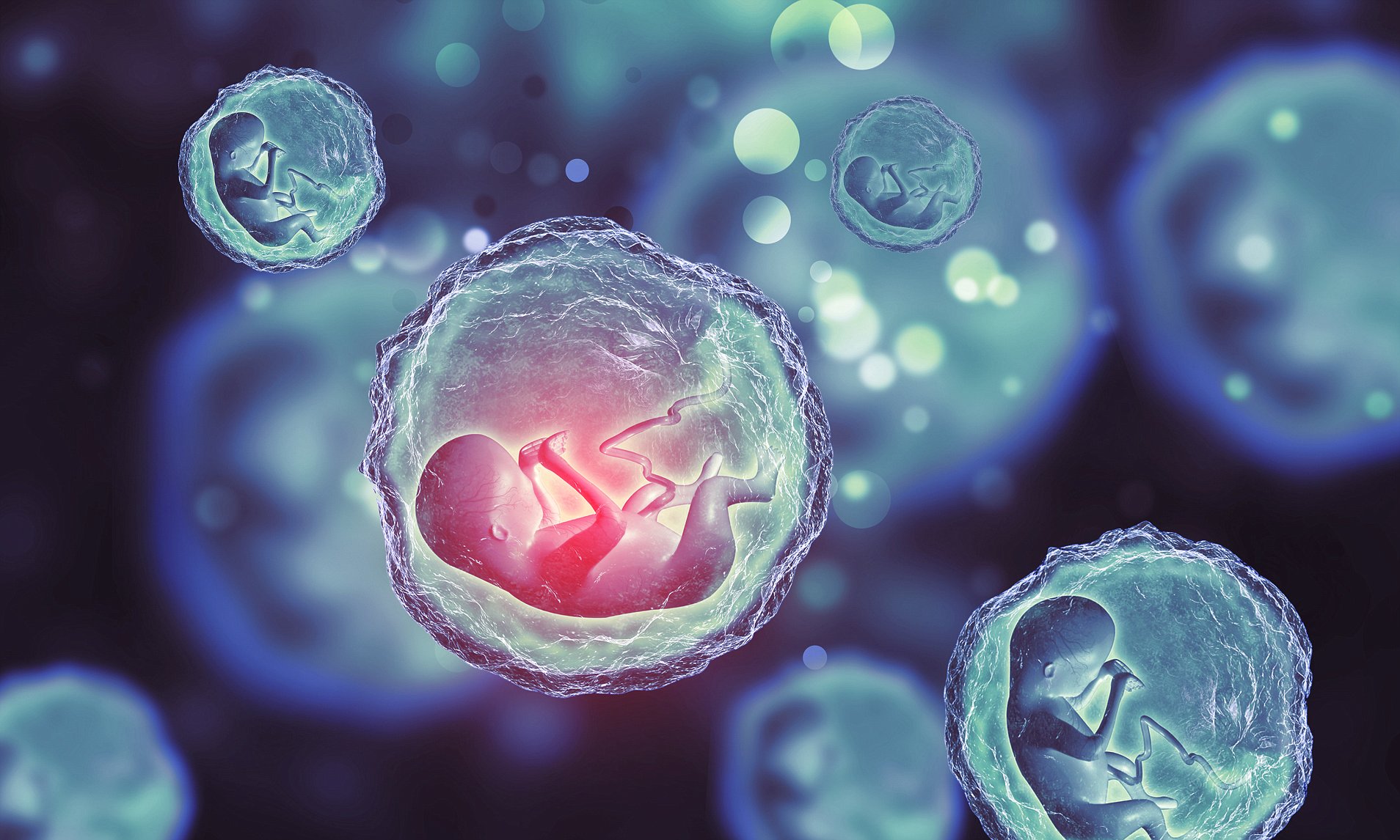
It created using stem cells looks like a classic example of a real 14-day-old embryo.
“embryo model” created
It even released hormones that made a pregnancy test in the lab positive.
The goal of embryo models is to provide an ethical framework for understanding the early stages of human life.
The initial few weeks after a sperm fertilises an egg are a time of tremendous change.
It’s from a collection of unclear cells to something that may be seen on a baby ultrasound.
This critical period is a major cause of miscarriage and birth abnormalities, yet it is little understood.
“It’s a black box, and that’s not a cliche – our knowledge is very limited,” Weizmann Institute of Science Prof Jacob Hanna tells me.
Starting Point
Embryo research is complicated legally, ethically, and technically.
However, there is currently a fast growing profession that mimics natural embryo development.
The Israeli team describes
The Israeli team describes this study, which was published in the journal Nature, as the first “complete” embryo model for imitating all of the main components that arise in the early embryo.
“This is a textbook image of a human day-14 embryo,” Prof Hanna adds, adding that “this hasn’t been done before.”
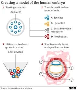
Instead of sperm and eggs, naive stem cells were used as the beginning material, and they were reprogrammed to have the ability to form any sort of tissue in the body.
Chemicals were then used to coax these stem cells into transforming into four different types of cells observed in the earliest stages of the human embryo:
- epiblast cells, which develop into the embryo (or foetus)
- Trophoblast cells develop into the placenta
- hypoblast cells, which develop into the yolk sac
- cells of extraembryonic mesoderm
The scientists took a step back and watched as 120 of these cells were combined in an exact ratio.
Arranging into a structure
Approximately 1% of the mixture began the process of spontaneously arranging itself into a structure that resembles, but is not identical to, a human embryo.
“I give great credit to the cells – you bring the right mix and have the right environment, and it just takes off,” Prof Hanna explains. “That’s an amazing phenomenon.”
The Embryo Models
The embryo models were allowed to grow and develop until they were comparable to a fertilised embryo 14 days later. This is the legal limit in several nations for normal embryo research.
Despite the late-night video conversation, I can hear Prof Hanna’s passion as she walks me through the “exquisitely fine architecture” of the embryo model.
I can see the trophoblast encircling the embryo, which would typically become the placenta. It also comprises the lacuna cavities, which fill with the mother’s blood to provide nutrients to the infant.
There is a yolk sac, which performs some of the functions of the liver and kidneys, as well as a bilaminar embryonic disc, which is one of the important features of this stage of embryo development.
‘Makes logic’
The objective is that embryo models will aid scientists in explaining how different types of cells form, witnessing the earliest stages of organ development, and understanding hereditary or genetic illnesses.
Other components of the embryo will not form until the early placenta cells can surround it, according to this study.
There is even talk of boosting IVF success rates by understanding why certain embryos fail or using the models to assess whether drugs are safe during pregnancy.
Prof Robin Lovell Badge of the Francis Crick Institute, who studies embryo development, tells me that these embryo models “look pretty good” and “look pretty normal.”
To read our blog on “Fermilab scientists are closing in on 5th force of nature,” click here